Technologies
Size Exclusion Chromatography (SEC) - IZON automated fraction collector (AFC)
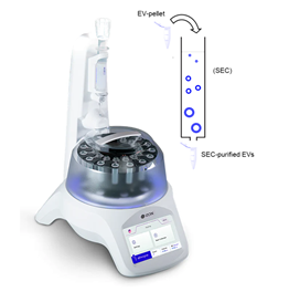
The qEV Automated fraction collector (AFC) system from IZON is designed for streamlined and reproducible size exclusion chromatography purification of extracellular vesicles (EVs) from biological samples. It automates the purification process using Izon's qEV isolation technology, ensuring high recovery and purity of EVs with minimal manual intervention. This system enhances throughput and consistency, making it ideal for large-scale research and clinical applications in EV-based diagnostics and therapeutics. Available columns can balance yield recovery and EV purity based on the chosen resin pore size (20, 35 and 70 nm). Colum sizes are additionally optimized based on sample volume (0.150 – 10 mL). Custom colums available upon request.
Tangential Flow Filtration (TFF) - Repligen KR2i TFF System
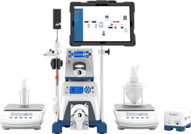
The KrosFlo® KR2i TFF System from Repligen is a fully automated tangential flow filtration (TFF) system designed for lab-scale purification and concentration of biomolecules. It features advanced control over key parameters like pressure, flow rate, and filtration performance, ensuring precise and reproducible results. Ideal for small-scale research, it supports a wide range of applications from cell therapy to protein and viral vector purification.
Western Blot - Bio-Rad
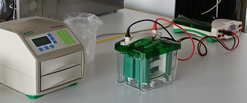
The EVR protein analysis workflow includes Bio-Rad’s Mini-PROTEAN® Tetra Vertical Electrophoresis Cell, paired with the PowerPac™ Basic Power Supply and Trans-Blot® Turbo™ Transfer System for rapid and reliable Western blotting. This system supports high-resolution polyacrylamide gel electrophoresis for protein separation, followed by efficient semi-dry transfer onto midi-format PVDF membranes. The streamlined design allows for quick gel casting, consistent voltage control, and rapid blotting—reducing transfer times to minutes without compromising protein integrity. Ideal for the analysis of EV-associated proteins, this platform enables reproducible results in targeted biomarker validation and downstream immunodetection assays.
Dynamic Light Scattering (DLS)
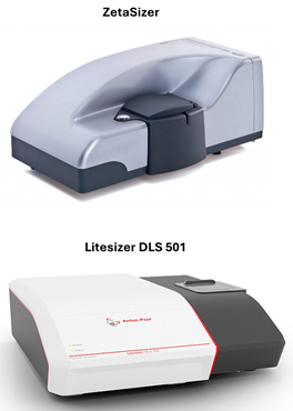
Dynamic Light Scattering (DLS), Photon Correlation Spectroscopy (PCS), or Quasi-Elastic Light Scattering (QELS) is a technique used to determine the size distribution profiles of small particles in suspension or emulsions. The analysis can be applied to micelles, polymers, proteins, colloids, and nanoparticles, including EVs. The EVR is equipped with a Zetasizer (Malvern), providing highly sensitive particle sizing and zeta potential measurements, ideal for stability studies and formulation development. The Litesizer DLS 501 (Anton Paar) can accurately measure particles from 0.3 nm to 12 µm. Measurements can be performed at three different detection angles, as well as in a Multi-Angle Particle Sizing (MAPS) mode for improved resolution and analytical precision. Determination of zeta potential uses a unique, patented cmPALS technology, for zeta potential measurement for particles ranging 1.3 nm to 100 µm in size. Litesizer DLS 501 can measure molecular mass and refractive index, and it can also be equipped with fluorescent and polarization filters. The Litesizer DLS 501 instrument is available upon request in collaboration with Prof. Paola Luciani's group.
Nanoparticle Tracking Analysis (NTA)
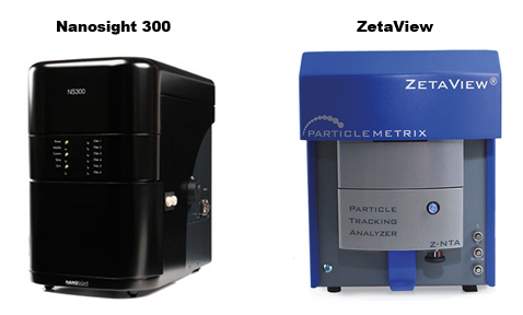
Nanoparticle Tracking Analysis (NTA) is a powerful technique for the high-resolution characterization of extracellular vesicles (EVs) and other nanoparticles in terms of yield and size distribution. Our facility is equipped with the NanoSight 300 (Malvern Panalytical) with 405, 488 and 531 nm laser modules. The multi-laser capability allows for fluorescence-based detection of labelled EV subpopulations. The ZetaView NTA system (Particle Metrix) enables high-resolution characterization of EVs and nanoparticles. Equipped with a 488 nm laser, zeta potential measurement capabilities, and an integrated cooling unit for enhanced stability, the system provides precise size distribution, concentration, and surface charge data. Its advanced multi-parameter analysis makes it an essential tool for studying EV heterogeneity, stability, and biomarker profiling. The ZetaView is available upon request in collaboration with Prof. Paola Luciani's group.
Interference Pattern - Videodrop (Myriade)
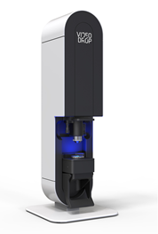
The Videodrop from Myriade is a label-free nanoparticle analysis system designed for rapid and real-time characterization of extracellular vesicles, viruses, and other nanoscale particles. Using interferometry, it provides direct, high-speed concentration and size distribution measurements without the need for fluorescent labelling. Its user-friendly interface and rapid analysis capabilities make it a powerful tool for quick sample assessments in EV research and bioproduction workflows.
Fluorescence Microfluidic Resistive Pulse Sensing (f-MRPS) - Spectradyne's ARC
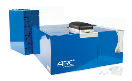
The ARC system from Spectradyne is an advanced microfluidic resistive pulse sensing (MRPS) instrument featuring dual-laser fluorescence (488 and 647 nm) detection for high-precision extracellular vesicle and nanoparticle characterization. Unlike traditional optical techniques, MRPS delivers accurate concentration and size distribution measurements across a broad dynamic range, while fluorescence capabilities enable the analysis of EV surface markers and cargo. This technology is ideal for robust, quantitative assessment of EV populations in biomarker discovery and therapeutic development.
Super Resolution Microscopy - ONI Nanoimager
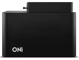
The ONI Nanoimager® is a cutting-edge super-resolution fluorescence microscope designed for single-molecule and extracellular vesicle (EV) imaging with nanometer precision. Equipped with four lasers and advanced optical modalities, it enables high-resolution studies of cells, but also EV structure, cargo, and interactions. The system supports multiple super-resolution techniques, including direct Stochastic Optical Reconstruction Microscopy (dSTORM) and Photoactivated Localization Microscopy (PALM), allowing for precise subcellular localization and molecular mapping. Single-particle tracking (SPT) provides real-time insights into EV dynamics, while DNA-PAINT enhances spatial resolution for molecular quantification. Additionally, single-molecule Förster Resonance Energy Transfer (smFRET) enables the study of molecular interactions within macromolecules at the 1-10 nm scale.
Collaborating Facilities
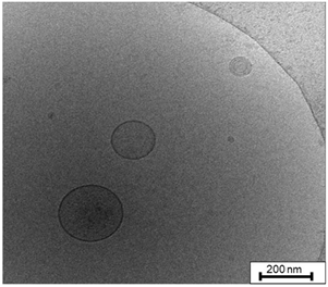
The EVR is actively collaborating with Core Facilities at the University of Bern, with SOPs and protocol development assistance for compatible downstream analyses with the Core Facility Proteomics and Mass Spectrometry (PMSCF) and the Flow Cytometry and Cell Sorting Facility (FCCS). Our team is also fully trained to perform Cryogenic electron microscopy (Cryo-EM), available upon request in collaboration with the Microscopy Imaging Center (MIC) at the University of Bern. (Image source: Zivko et al, Commun Biol. 2022.)